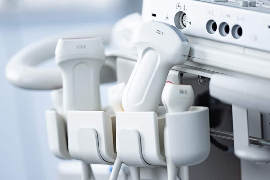
Ultrasounds or sonography is a common diagnostic exam medical professionals perform to assess the body's structure and organs. It uses high-intensity sound waves to create fetus imaging (in a pregnant woman), tissues, blood vessels, and organs. The sound waves produced from an ultrasound scanning machine bounce off the body parts' fluid and create an image.
Ultrasound techniques have evolved, and medical professionals use them to make the diagnoses. It helps medical experts view the internal organs of the body as they perform their function. An ultrasound procedure can examine the body parts, including the abdomen, female pelvis, breasts, prostate, thyroid, parathyroid, scrotum, and vascular system. During pregnancy, doctors perform several ultrasounds to evaluate fetus development.
Let’s delve into the details of the different types of ultrasounds and how doctors use them.
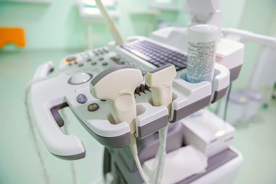
An abdominal ultrasound examines the internal organs that may include the spleen, gall bladder, liver, kidneys, bladder, and pancreas. This ultrasound scan helps medical professionals diagnose various conditions and evaluate the damage caused by different ailments and illnesses. As an abdominal ultrasound provides doctors a real-time image, they use it for;
Pelvic ultrasound is the most well-known form of ultrasound scanning you may undergo in pregnancy. It is typically an imaging test gynecologists use to monitor the growing baby in the mother’s womb. Apart from maternity use, a pelvic ultrasound can also examine ovaries, bladders, uterus, and prostate gland. Doctors may use to diagnose condition like;
Patients undergoing a transabdominal ultrasound have a full urinary bladder scan. They need to lie on the back and apply a gel to the abdomen. An ultrasound expert rubs over transducer over the examination area as it releases high-frequency sound waves. Like other ultrasounds, the procedure of Transabdominal is straightforward.

As the name reflects, transvaginal ultrasound scans the bladder through the vagina. Women need to empty their bladders before scanning to help a gynecologist examine a clear image. They also lie face up on the back with their legs stirrup.
An ultrasound technician inserts the transducer into the vagina for the test. The transducer's size in transvaginal ultrasound is smaller compared to a standard speculum in Pap tests. Technicians use a protective gel and cover for lubrication on the transducer before insertion. They insert the first few inches of a transducer in the vagina and move it in a circular motion to get the images.
Most women receive a transvaginal ultrasound for the diagnosis of pelvic pain. The scanning test is more comfortable and easy than a manual gynecologic test.
Transrectal ultrasound is for examining the prostate gland. The doctor or an ultrasound technician inserts the transducer through the rectum and allows sound waves to reach the prostate.
Like other inserted scanning process, technicians use a protective gel and cover for lubrication on the transducer before insertion. They move around the transducer to obtain rectum images from multiple angles. For the scanning test, a patient needs to lie down on the left side and bend up the knees toward the chest.
If doctors find a lesion during an ultrasound exam, they send it for biopsy. The ultrasound images help radiologists guide the needles to the prostate gland and sample enlarged or abnormal tissues.
Obstetric ultrasound (OB scanning) refers to the focused use of high-frequency waves to evaluate a pregnant woman’s condition and fetus or embryo. Doctors recommend this ultrasound on the clinical requirements, which may include;
Women need to lie on their back and expose their abdominal area for an ultrasound scanning. The obstetric ultrasound exam takes about 35-45 minutes. It is a non-invasive procedure, but there can be varying degrees of uneasiness and discomfort due to transducer pressure.
An ultrasound technician presses the transducer firmly over the abdomen to get closer to the fetus and better see the structure. The discomfort is for a few minutes only.
Ultrasound scanning of the carotid aorta system provides a quick, non-invasive means of spotting blockage of blood flow in your neck arteries. The arteries go to the brain, and even a minor blockage may produce a mini-stroke.
Doctors use abdominal aorta ultrasound to evaluate conditions like an aneurysm. It is an abnormal enlargement of the carotid aorta from atherosclerotic disease.
Ultrasound imaging or scanning of the breast aims to evaluate the internal structure of the breast. It uses waves to create pictures and primarily helps doctors to diagnose lumps and abnormalities in the breast.
Typically, doctors perform a breast ultrasound scanning when they find a lump in the breast during a mammogram, physical exam, or breast MRI. The procedure is non-invasive, safe, and doesn’t use radiations. The process requires no special arrangement. Women need to undress up to the waist and wear a loose gown during the ultrasound.
You may undergo different types of ultrasound procedures in a nutshell, depending on the body part your doctor needs to examine. There can be internal and external ultrasound scans. Thus, the article includes information on the most common ultrasound scans.
If you have any questions, concerns, apprehensions, unease, or worry about your fetus’ development contact your health care provider immediately.


ALL WARRANTIES OF ANY KIND WHATSOEVER EXPRESS, IMPLIED, AND STATUTORY, ARE HEREBY DISCLAIMED. ALL IMPLIED WARRANTIES OF MERCHANTABILITY AND FITNESS FOR A PARTICULAR PURPOSE ARE HEREBY DISCLAIMED. THE PRODUCTS SOLD, INCLUDING SONOGRAMS, ULTRASOUNDS, FAKE PREGNANCY DOCUMENTS, AND FAKE PREGNANCY TESTS ARE SOLD ‘AS IS’ BASIS.
THE SITE CANNOT AND DOES NOT CONTAIN [MEDICAL/ LEGAL/ FITNESS/ HEALTH/ OTHER] ADVICE. THE INFORMATION IS PROVIDED FOR PRANKS PURPOSES ONLY AND IS NOT A SUBSTITUTE FOR PROFESSIONAL ADVICE.
ACCORDINGLY, BEFORE TAKING ANY ACTIONS BASED UPON SUCH INFORMATION, WE ENCOURAGE YOU TO CONSULT WITH THE APPROPRIATE PROFESSIONALS. WE DO NOT PROVIDE ANY KIND OF MEDICAL/ LEGAL/ FITNESS/ HEALTH ADVICE. THE USE OR RELIANCE OF ANY INFORMATION CONTAINED ON THIS SITE, OR OUR MOBILE APPLICATION, IS SOLELY AT YOUR OWN RISK.
THIS WEBSITE DOES NOT PROVIDE MEDICAL ADVICE. THE INFORMATION, INCLUDING BUT NOT LIMITED TO, TEXT, GRAPHICS, IMAGES AND OTHER MATERIAL CONTAINED ON THIS WEBSITE ARE FOR PRANK PURPOSES ONLY. NO MATERIAL ON THIS SITE IS INTENDED TO BE A SUBSTITUTE FOR PROFESSIONAL MEDICAL ADVICE, DIAGNOSIS OR TREATMENT. ALWAYS SEEK THE ADVICE OF YOUR PHYSICIAN OR OTHER QUALIFIED HEALTH CARE PROVIDER WITH ANY QUESTIONS YOU MAY HAVE REGARDING A MEDICAL CONDITION OR TREATMENT AND BEFORE UNDERTAKING NEW HEALTH CARE REGIMEN, AND NEVER DISREGARD PROFESSIONAL MEDICAL ADVICE OR DELAY IN SEEKING IT BECAUSE OF SOMETHING YOU HAVE READ ON THIS WEBSITE.
THE PARTIES AGREE THAT ANY PRODUCT PURCHASED ON THE BABY MAYBE WEBSITE SHALL NOT BE USED FOR ANY PROPOSE OTHER THAN AS A PRANK. WITHOUT EXCEPTION NO BABY MAYBE PRODUCT SHALL BE PROVIDED/SUBMITTED TO ANY GOVERNMENTAL OR OTHER AGENCY, MEDICAL DOCTOR, ARBITER OF A DISPUTE, AS PROOF OF PREGNANCY, PAST OR CURRENT, OR TO CLAIM ANY BENEFIT FOR WHICH A PREGNANT WOMAN MAY BE ELIGIBLE, OR ENTITLED TO RECEIVE, BASED ON HER BEING PREGNANT. NO HIPAA PROTECTED PATIENT HEALTH INFORMATION CONNECTED TO ANY BABY MAYBE PRODUCT, IS INTENDED, OR CONVEYED, WITH RESPECT TO THIS SALE.
THE PARTIES AGREE THAT BABYMAYBE IS NOT RESPONSIBLE FOR ANY LIABILITY WHATSOEVER FOR DELAYS IN SHIPPING THE PRODUCT. THE PARTIES FURTHER AGREE THAT THE SOLE REMEDY FOR ANY SHIPPING DELAYS IS THE REFUND OF THE PURCHASER’S PAYMENT FOR THE PRODUCT.
THE PARTIES AGREE THAT THE FORUM FOR ANY LEGAL ACTION ASSOCIATED WITH THE SALE AND PURCHASE OF THE PRODUCT IS THE STATE OF ILLINOIS.