An ultrasound, whether it's during pregnancy or other medical conditions, provide valuable information to doctors. They are an integral and standard part of medical care, particularly for pregnant women. An ultrasound report result allows doctors to monitor the growth of the baby and detect complications and abnormalities.
They are also vital for predicting the due date and determining whether a pregnant woman is carrying multiples. Plus, gynecologists use ultrasound to see and assess the placenta's position and the gender of the baby.
But how often can you get an ultrasound or how many scans are safe to take during pregnancy are important questions to consider. If you're expecting a baby and have these questions in mind, we have the valuable information to find the answers.
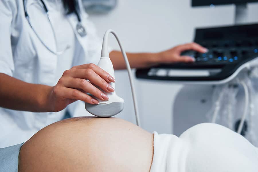
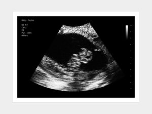
Ultrasound is an imaging or scanning technology. It uses resonance and sound waves to produce a bounce in the womb's fluid and create images. An ultrasound technician or a doctor places a wand (transducer) in the vagina or belly to emit sound waves.
The echo of waves typically produces movements in the bones, tissues, and fluid of the baby. Then, it picks up the waves and echoes to translate an image of the fetus parents can see on the computer screen.
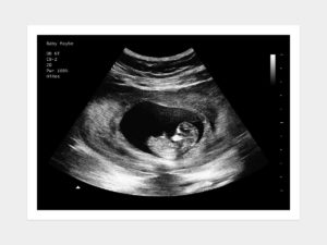
During early pregnancy, the ultrasound scan confirms pregnancy location and the fetal heartbeat. However, in later stages, the sonography reveals placenta location and fetal growth. It also shows the general anatomy and health of the baby.
In the final days of pregnancy, sonography or ultrasound scans are very useful to assess the progress and cervix length. It is important as doctors need to confirm that you're not in preterm labor. Final stage ultrasounds also verify if the fetus is in the right position (head-down), which is one of the major requirements for vaginal delivery.
Keep in mind that an ultrasound scan, regardless of its stage, doesn't hurt. You might feel a little messy or cold when the transducer touches the belly because of the gel on it. To-be mommies should wear two-piece clothes to allow quick and easy access to the tummy.
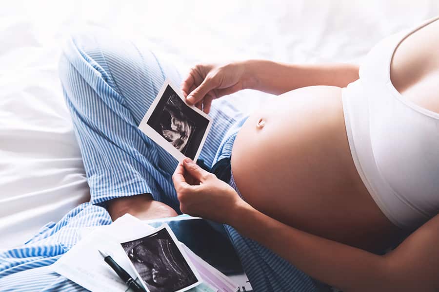
You may also not see a clear image of the baby during the initial phases of pregnancy in an ultrasound report. It gets clearer overtime, usually around 12 to 13 weeks, making it an ideal time to reveal your loved ones' exciting news.
The number of times you need to do an ultrasound may vary depending on how complicated your pregnancy is. Here is an overview of typical ultrasound scans you may undergo while expecting.
Generally, you may have your first ultrasound or sonogram after 6 to 8 weeks of your pregnancy. Many doctors consider conducting this test only if there is any risk in your pregnancy. These may include your history of miscarriages, defects, abdominal pain, and bleeding.
Your doctor may conduct the first ultrasound transvaginally to obtain a clearer picture of the fetus. If this is the case, the doctor will place a wand-like transducer to transmit high-frequency waves through the uterus. The sound waves send signals to the machine to make reflections of an image of the fetus.
If it is a six-week pregnancy ultrasound, it is possible to listen to the baby's heartbeat. Your practitioner can also track milestones, predict the due date, and find the fetus's number in the womb. At six weeks, doctors can also whether your pregnancy is ectopic.
You may undergo a "dating ultrasound" if you skip the early pregnancy scan. It takes place around 8-13 weeks of pregnancy. The sonography scan provides the same information as due date, "crown-rump length" of the baby (measurement), fetal heartbeat, and babies' number.
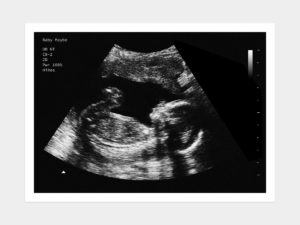
This is a detailed ultrasound that generally takes place between 18 to 20 weeks of pregnancy. It usually lasts up to 20 to 50 minutes if you're pregnant with one baby. If you're going to have twins, triplets, or quadruplets, the ultrasound may take a little longer.
At this stage, doctors do a thorough checkup of the baby by assessing his/her heart rate and abnormalities in the brain, kidneys, livers, and heart. Doctors also count the baby's toes and fingers, examine the placenta, and measure amniotic fluid levels.
It is worth mentioning that ultrasounds are safe for t baby and mother when conducted for medical purposes.

Although ultrasound scanning requires no radiation, only professional technicians who specialize in interpreting the results accurately should conduct it. Preferably, an ultrasound technician has a certificate in obstetrical ultrasound from the American Institute of Ultrasound in Medicine.
Moreover, ultrasound scans are safe to conduct but only in a medical setting as they may heat the tissues. Sometimes, an ultrasound machine produces bubbles or cavitations during the test.
According to experts, the heat and cavitations might have a long-term effect on the baby, especially when performed without a medical need. Therefore, medical professionals emphasize performing a sonography or ultrasound scan only when a patient requires it for a medical purpose based on a doctor's prescription. Also, you need to make sure that only a trained professional performs it.
While many women expect at least three ultrasound scans during pregnancy in the first two trimesters, there are several reasons doctors prescribe for more. Ultrasounds are crucial when monitoring the growth of the baby. It is normal to become anxious with the idea of undergoing more screenings or conducting tests beyond your routine checkups.
The ultrasound scans are safe for your health, and you can get them 5 to 6 times during pregnancy as per your doctor's recommendation. However, make sure you discuss unforeseen issues pertaining to your pregnancy and the effects of an ultrasound scan on the baby.
If you have any questions, concerns, apprehensions, unease, or worry about your fetus’ development contact your health care provider immediately.


ALL WARRANTIES OF ANY KIND WHATSOEVER EXPRESS, IMPLIED, AND STATUTORY, ARE HEREBY DISCLAIMED. ALL IMPLIED WARRANTIES OF MERCHANTABILITY AND FITNESS FOR A PARTICULAR PURPOSE ARE HEREBY DISCLAIMED. THE PRODUCTS SOLD, INCLUDING SONOGRAMS, ULTRASOUNDS, FAKE PREGNANCY DOCUMENTS, AND FAKE PREGNANCY TESTS ARE SOLD ‘AS IS’ BASIS.
THE SITE CANNOT AND DOES NOT CONTAIN [MEDICAL/ LEGAL/ FITNESS/ HEALTH/ OTHER] ADVICE. THE INFORMATION IS PROVIDED FOR PRANKS PURPOSES ONLY AND IS NOT A SUBSTITUTE FOR PROFESSIONAL ADVICE.
ACCORDINGLY, BEFORE TAKING ANY ACTIONS BASED UPON SUCH INFORMATION, WE ENCOURAGE YOU TO CONSULT WITH THE APPROPRIATE PROFESSIONALS. WE DO NOT PROVIDE ANY KIND OF MEDICAL/ LEGAL/ FITNESS/ HEALTH ADVICE. THE USE OR RELIANCE OF ANY INFORMATION CONTAINED ON THIS SITE, OR OUR MOBILE APPLICATION, IS SOLELY AT YOUR OWN RISK.
THIS WEBSITE DOES NOT PROVIDE MEDICAL ADVICE. THE INFORMATION, INCLUDING BUT NOT LIMITED TO, TEXT, GRAPHICS, IMAGES AND OTHER MATERIAL CONTAINED ON THIS WEBSITE ARE FOR PRANK PURPOSES ONLY. NO MATERIAL ON THIS SITE IS INTENDED TO BE A SUBSTITUTE FOR PROFESSIONAL MEDICAL ADVICE, DIAGNOSIS OR TREATMENT. ALWAYS SEEK THE ADVICE OF YOUR PHYSICIAN OR OTHER QUALIFIED HEALTH CARE PROVIDER WITH ANY QUESTIONS YOU MAY HAVE REGARDING A MEDICAL CONDITION OR TREATMENT AND BEFORE UNDERTAKING NEW HEALTH CARE REGIMEN, AND NEVER DISREGARD PROFESSIONAL MEDICAL ADVICE OR DELAY IN SEEKING IT BECAUSE OF SOMETHING YOU HAVE READ ON THIS WEBSITE.
THE PARTIES AGREE THAT ANY PRODUCT PURCHASED ON THE BABY MAYBE WEBSITE SHALL NOT BE USED FOR ANY PROPOSE OTHER THAN AS A PRANK. WITHOUT EXCEPTION NO BABY MAYBE PRODUCT SHALL BE PROVIDED/SUBMITTED TO ANY GOVERNMENTAL OR OTHER AGENCY, MEDICAL DOCTOR, ARBITER OF A DISPUTE, AS PROOF OF PREGNANCY, PAST OR CURRENT, OR TO CLAIM ANY BENEFIT FOR WHICH A PREGNANT WOMAN MAY BE ELIGIBLE, OR ENTITLED TO RECEIVE, BASED ON HER BEING PREGNANT. NO HIPAA PROTECTED PATIENT HEALTH INFORMATION CONNECTED TO ANY BABY MAYBE PRODUCT, IS INTENDED, OR CONVEYED, WITH RESPECT TO THIS SALE.
THE PARTIES AGREE THAT BABYMAYBE IS NOT RESPONSIBLE FOR ANY LIABILITY WHATSOEVER FOR DELAYS IN SHIPPING THE PRODUCT. THE PARTIES FURTHER AGREE THAT THE SOLE REMEDY FOR ANY SHIPPING DELAYS IS THE REFUND OF THE PURCHASER’S PAYMENT FOR THE PRODUCT.
THE PARTIES AGREE THAT THE FORUM FOR ANY LEGAL ACTION ASSOCIATED WITH THE SALE AND PURCHASE OF THE PRODUCT IS THE STATE OF ILLINOIS.